Contact





Knee Joint Surgery

Surgery is performed when improvement is difficult to achieve with conservative treatment (medication and physical therapy). The main types include surgery to change the shape of the knee joint, joint replacement, and joint fusion.
-
Arthroscopic Meniscus Repair
For a damaged meniscus, suturing and smoothing of uneven cartilage are performed through small incisions. A damaged meniscus causes swelling and pain during daily activities and sports. If left untreated, it can lead to knee osteoarthritis over the course of about ten years, so it should be repaired with sutures whenever possible. A wide variety of instruments are available for arthroscopic surgery, and mastering them is where a knee surgeon truly demonstrates their skill.
-
Total Knee Arthroplasty (TKA)
This surgery thinly resurfaces the deformed surfaces of the knee joint and replaces them with an artificial joint. Because the painful areas are covered with metal, it is highly effective in relieving knee pain from soon after surgery. During the operation, an incision is made in the front of the knee, and the damaged parts of the femur and tibia (and patella) are removed. The bone is shaped to fit the metal components, and the artificial joint is fixed to the bone with an adhesive called bone cement to complete the surgery. The patella (kneecap) is also replaced with polyethylene as needed. Recently, new technologies such as navigation systems have been developed, allowing surgery to be performed with greater precision. There are two types of knee arthroplasty: total knee arthroplasty and unicompartmental knee arthroplasty. Total knee arthroplasty is performed when the entire knee joint is damaged, while unicompartmental knee arthroplasty, which partially replaces the joint, is performed when only the inner side is damaged. Unicompartmental knee arthroplasty offers faster postoperative recovery, better knee flexion, and less discomfort.
-
Medial Opening-Wedge High Tibial Osteotomy (DTO) / High Tibial Osteotomy (HTO)
Because deformity places excessive stress on the inner side of the knee, this surgery cuts the patient's own bone to correct the bow-legged alignment and shift the load to the relatively healthy cartilage on the outer side. This surgery changes the leg alignment from bow-legged to knock-kneed. It preserves or regenerates the patient's own knee, making it possible to sit in the traditional kneeling position (seiza) and return to sports or work such as farming. However, it takes three to four months after surgery for the bone to heal, so thorough rehabilitation is essential.
Spine and Spinal Cord Surgery
Keitaro Matsukawa, Director, Spine and Spinal Cord Center
-
Treatment of Lumbar Spine Disorders with a New Spinal Endoscopic Surgery (UBE)
In recent years, advances in endoscopic technology have made minimally invasive spine surgery, which minimizes the burden on the body, widely available. Our clinic has introduced Unilateral Biportal Endoscopy (UBE), a type of spinal endoscopic surgery, to treat lumbar spinal canal stenosis (a condition in which the passage for the nerves becomes narrow) and lumbar disc herniation.
-
How It Differs from Conventional Surgery
In conventional surgery, an incision of about 5 cm is made in the skin, and the muscles attached to the spine are detached to relieve pressure on the nerves. In UBE, by contrast, two small incisions of 5–8 mm are made, and surgery is performed through the gaps between the muscles. An endoscope (camera) is inserted through one opening and surgical instruments through the other, allowing the surgeon to operate while viewing the surgical field. This method makes it possible to proceed with surgery while observing the nerves and surrounding tissues under magnification and minimizing damage to the muscles.
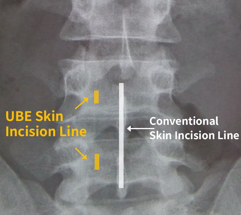

UBE Surgery Through Two Small Incisions
An endoscope is inserted through one incision and surgical instruments through the other to perform the procedure.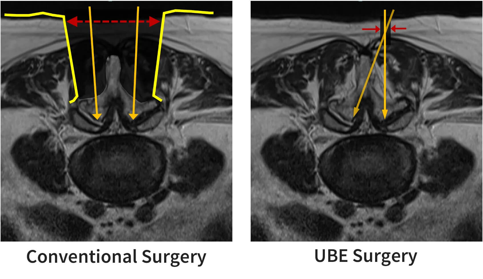
Comparison of Conventional Surgery and UBE Surgery
While conventional surgery requires extensive detachment of the muscles around the spine, UBE treats the area around the nerves through small incisions and the gaps between the muscles.The greatest advantage of this surgery is that the two incisions provide separate pathways for viewing and for working. This greatly increases freedom of surgical maneuvering and broadens its applicability to a variety of conditions, including lumbar disc herniation, lumbar spinal canal stenosis, and degenerative lumbar spondylolisthesis.
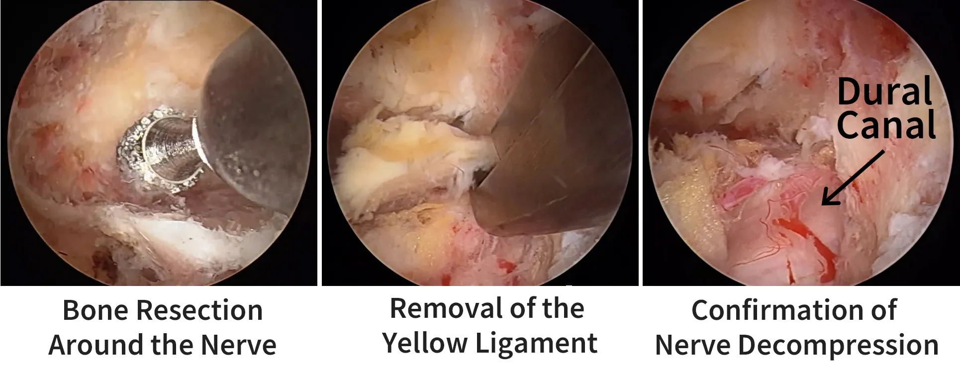
Representative Case of UBE Surgery for Lumbar Spinal Canal Stenosis
The endoscopic image is displayed on a monitor, and the surgery proceeds while the area is examined in detail. The surgery is completed after confirming that the environment around the nerves has been sufficiently improved.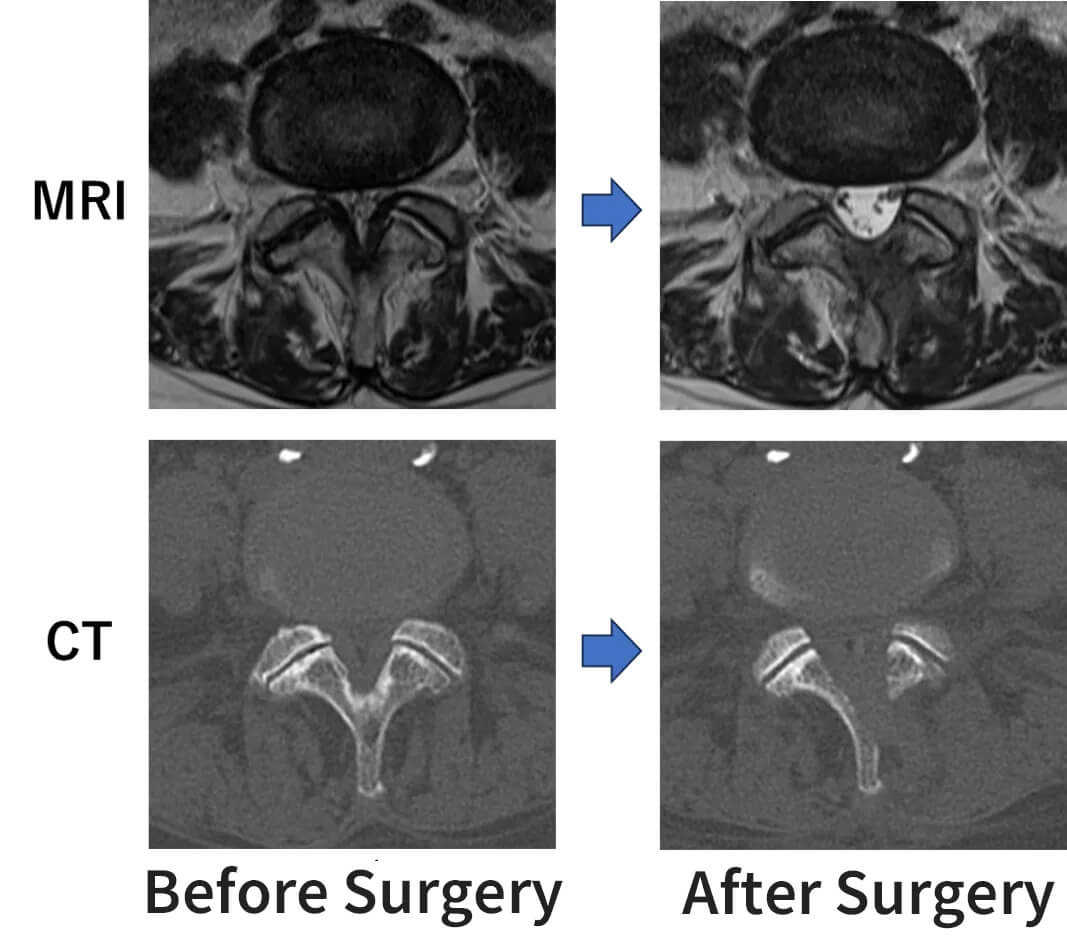
Comparison of Preoperative and Postoperative MRI and CT Images
With minimal bone removal, the passage for the nerves has been appropriately widened. -
Treatment of Lateral Lesions (Foraminal Lesions)
Some lumbar spine disorders involve “lateral lesions” (such as lateral disc herniation and foraminal stenosis), in which the nerves are compressed on the outer side of the spine (the foramen). Because this area lies near the facet joints, which contribute to spinal stability, conventional surgery may require extensive bone removal, and as a result, spinal fusion (surgery using screws) may be considered.
With UBE, however, it is possible to approach the lesion through the most appropriate route according to its location and condition, improving the environment around the nerves while limiting the extent of bone removal. “Another hospital told me I need surgery with screws. Can it be treated without them?” This is a question we often hear from our patients. Of course, careful judgment based on each condition is required, but even for conditions where spinal fusion would have been considered with conventional methods, endoscopic treatment such as UBE may make it possible to avoid fusion. -
What Is the Ideal Spine Surgery?
Depending on the condition, a more invasive procedure is sometimes unavoidable. However, if the same results can be achieved for the same condition, we want to reduce the burden on the body as much as possible. Of course, a smaller incision is not always better in itself. Each patient has different wishes and values. What matters is “diversity in surgical treatment.” We believe our mission is to provide treatment that meets each patient's wishes. At our clinic, we consider tailor-made surgery based on each patient's condition, including for patients who wish to receive minimally invasive treatment. We continually refine our skills to provide the best possible treatment, looking not only at the present but also five and ten years ahead.
“Provide the patient in front of us with the surgery we would truly want to receive ourselves.”
This is our work as professionals.
Endoscopic Lumbar Interbody Fusion Using UBE
Keitaro Matsukawa, Director, Spine and Spinal Cord Center
-
Decompression and Fusion Surgery
Spinal surgery is broadly divided into two categories: “decompression surgery” and “fusion surgery.” Decompression surgery is a procedure designed to improve the environment around the nerves by removing bone from the “vertebral arch”—the bony structure that forms the roof over the nerves—in order to widen the spinal canal, through which the compressed nerves pass. The primary indication for this procedure is lumbar spinal stenosis.
On the other hand, “spinal fusion” is a surgical procedure performed not only to relieve nerve compression but also to correct and stabilize the alignment of the spine in cases of spinal instability or overall spinal deformity. The main indications for this procedure include lumbar spondylolisthesis, lumbar spondylolysis, and scoliosis or kyphosis. Generally, a fixation device called a pedicle screw is required, which is inserted into the spine through an incision in the back.
-
What Is Endoscopic Lumbar Interbody Fusion?
Lumbar interbody fusion is a surgical procedure in which damaged intervertebral disc tissue between the lumbar vertebrae is removed and replaced with bone harvested during the operation or an artificial implant called an interbody cage.
At our clinic, we also use the aforementioned UBE (Unilateral Biportal Endoscopy) for lumbar interbody fusion.
Compared to conventional fusion procedures that involve a large incision in the back, this method not only requires a smaller skin incision but also minimizes muscle damage. Its advantages include reduced postoperative pain, a shorter hospital stay, and a faster return to daily activities.Comparison of Skin Incisions Between Conventional Fusion and UBE Fusion
-
Details of the Surgical Procedure
Four small incisions are made on either side of the body to insert screws percutaneously. Through these incisions, an endoscope (camera) and surgical instruments are inserted between the muscles to perform nerve decompression and interbody procedures. Finally, screws are placed to stabilize the lumbar spine. Patients are generally able to walk the day after surgery and require a hospital stay of approximately 5–7 days. As with conventional fusion surgery, patients are required to wear a brace for 3–4 months postoperatively and should refrain from activities that place strain on the lower back for 6 months following surgery.
UBE Fusion in Practice
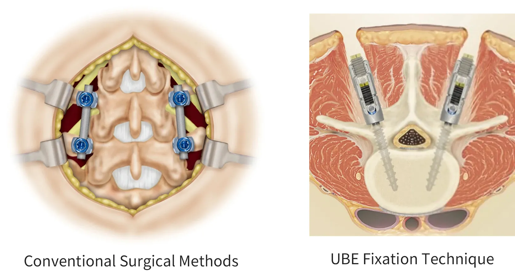
While conventional surgical methods required a large incision in the affected area, the UBE fusion procedure involves making a small incision and performing endoscopic manipulation and screw placement through the space between the muscles, thereby significantly reducing damage to the muscles.
-
Precautions
This procedure may not be indicated in cases of severe nerve compression or significant lumbar spinal deformity or osteoporotic changes. Additionally, if the amount of bone harvested for intervertebral bone grafting is insufficient, bone may be harvested from the pelvis via a small incision.
-
Representative Case (76-year-old woman)
The patient presented with walking difficulties due to pain in both lower limbs. As walking even 200 meters had become difficult, the patient was diagnosed with degenerative spondylolisthesis of the fourth lumbar vertebra and underwent endoscopic lumbar interbody fusion using the UBE system. After surgery, the lower-limb symptoms resolved. Postoperative MRI confirms that the spinal canal has widened.